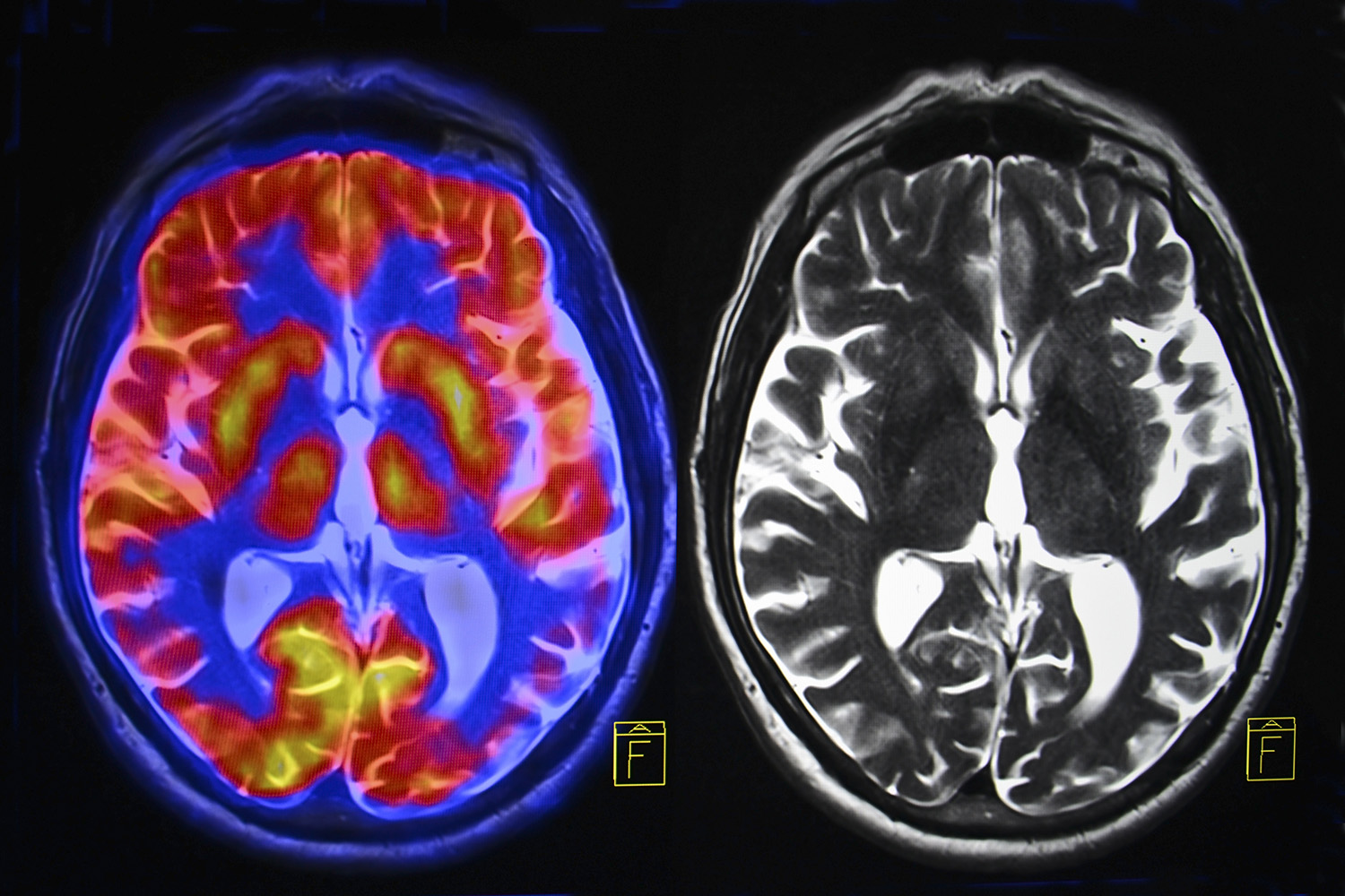
It has been known for more than two decades that elevated cholesterol was associated with increased risk for Alzheimer’s Disease (AD).[1] It is also known that the ApoE gene produces a protein that transports fats, including cholesterol, into brain cells.
In the human population, there are three variants of the ApoE gene. Seven percent of the population has ApoE2, which confers increased risk for atherosclerosis; 79% has ApoE3, which confers no disease risk; and 14% of the population has the ApoE4 variant, which increases the risk for AD.
But, not everyone with either the ApoE4 gene or elevated cholesterol gets AD. Could there be an interaction between the higher concentrations of cholesterol and a specific ApoE gene variant that does increase the risk? The answer seems to be yes. Research has demonstrated that modulation in cholesterol alters the ApoE gene activity.[2] Further research has discovered a nexus between these two factors. A high-fat diet was demonstrated to increase insulin resistance and cognitive decline in all groups, whether ApoE4 or ApoE3. However, those with the ApoE4 gene had “exaggerated” deficits in the part of the brain specific to new learning and forming new memories in the hippocampus, but when those with the ApoE4 gene were placed on a low-fat diet for one month, all deficits reversed and learning and cognitive function returned to normal![3] The researchers concluded that those with two copies of the ApoE4 gene were particularly susceptible to neuronal and cognitive impairments due to insulin resistance caused by a high-fat diet.
Having demonstrated that low-fat diets result in improved cholesterol profiles and subsequent improvement in brain and cognitive function — especially in those with two copies of the ApoE4 gene, researchers examined whether cholesterol lowering medications could offer the same benefit. Data from long term clinical trials have demonstrated that some, but not all, cholesterol lowering medications conferred reduced risk of AD and better cognitive performance, especially in those with two copies of the at-risk ApoE4 gene. The greatest positive effect was seen with atorvastatin (P = .026) and the least with lovastatin (no significant difference found). Those individuals with two copies of the at-risk gene and who already had symptoms of AD, but received statin medication, had significantly better cognitive function over the course of a 10-year follow-up, compared with those who did not receive the statins (P < .01).[4]
Recently, researchers from Johns Hopkins University have discovered another brain protein that appears to be involved and works in concert with elevated b-amyloid to cause the cognitive and memory impairments of AD. The NPTX2 gene is one of the first genes to get activated when new memories are forming. If you are trying to remember what you are reading in this article, then normally NPTX2 would activate and produce the protein with the same name (NPTX2). This protein acts as an instigator and activator of synaptic signaling and neural circuit recruitment, critical in the formation of new memories. Without this protein, the neural circuits cannot effectively synchronize to form new memories. When this gene is turned down at the same time b-amyloid is building up in the brain, the neural circuits’ ability to adapt and organize is impaired, contributing to the cognitive and memory decline of AD. Individuals with high b-amyloid and high NPTX2 did not show cognitive changes of AD, and individuals with low NPTX2 and low b-amyloid also did not show impairment of cognition and memory. This study documented that both high b-amyloid and low NPTX2 were required for the negative outcomes. The good news is that the cause of suppressing NPTX2 is different than what causes elevations in b-amyloid.[5] This provides additional opportunities to make lifestyle changes to protect our brains and prevent dementia — even if one has the at-risk genes.
So, what turns on the NPTX2 gene? Activity of the neurons themselves![6] Staying mentally engaged and cognitively active — people who are lifelong learners — keep the neurons active and NPTX2 turned on, with reduced risk of AD. Additionally, externally firing the neurons with treatments such as electroconvulsive therapy has been documented to increase the expression of this gene.[7] These two findings makes it likely that any activity that increases the neuronal firing will activate the NPTX2 gene and may be one of the benefits of transcranial magnetic stimulation, which causes neuronal firing via magnetic waves rather than electrical pulses.
Not only does NPTX2 enhance learning, neural circuitry recruitment, synchronicity, and brain neural plasticity, it also modulates a receptor (AMPA) involved in non-programmed cell death. Therefore, while normal amounts of NPTX2 are neural protective, and low amounts increases the risk of dementia, significantly higher than normal activity of NPTX2 can trigger AMPA and instigate unscheduled cell death. This, unfortunately, appears to occur in persons with Parkinson’s disease and Lewy Body dementia, where NPTX2 is increased by more than 800% in the motor pathways.[8]
Another protein critical in maintaining brain health is repressor element 1-silencing transcription (REST) factor. REST functions within the cell like a conductor of an orchestra, directing various genes to sound out (express themselves) or be silent (turn off). As a result, REST is involved in determining how neurons develop, what function they fulfill, their connections and networking to other neurons, and, as expected, is highly active in childhood during the massive remodeling of brain development.
In the past, it was believed REST became inactive after a person reached adulthood. However, recent research has discovered REST is active in older brains and functions to protect the memory circuits (hippocampus) from damage due to hyperexcitation and plays a key role in protecting the brain from damage associated with aging. Reduced levels of REST are associated with loss of brain volume in the hippocampus (memory circuits) and increased cognitive impairment. In persons who have the toxic build up of protein associated with Alzheimer’s dementia (amyloid and tau), those with high REST activity did not demonstrate cognitive decline or progress to dementia, supporting the idea that REST is neural protective. The critical question: What affects the availability of REST? Chronic mental stress suppresses REST, contributing to accelerated aging and cognitive decline, whereas meditation, that reduced stress, is associated with increased levels of REST and subsequent brain health. [9]
With all of this in mind, genetics appears to account for about one-third of the risk of developing AD. What is the key then that contributes to developing AD, if it isn’t simply genetics? Strong evidence points to inflammation, which contributes to insulin resistance in the brain, that causes a cascade of events, resulting in the death of brain cells and the development of AD. Exercise, along with most of the other modifiable factors (sufficient sleep, anti-inflammatory diet, stress management, etc.), reduces inflammation and insulin resistance, keeps neurotrophins (proteins that act like fertilizer for the neurons), REST, NPTX2, and other protective factors turned on, thereby preventing the development of AD.
While aging is inevitable, dementia is not! We can make choices to protect our brains and prevent the development of late-life Alzheimer’s dementia. I recommend my new book, The Aging Brain: Proven Steps to Prevent Dementia and Sharpen Your Mind, which is an integrative examination of the various contributing factors to AD and outlines a comprehensive action plan to slow the aging process and keep our brain healthy.
[1] Jarvik GP, et al. Interactions of apolipoprotein E genotype, total cholesterol level, age, and sex in prediction of Alzheimer’s disease: a case-control study. Neurology. 1995;45(6):1092–6.
[2] Petanceska SS, et al. Changes in apolipoprotein E expression in response to dietary and pharmacological modulation of cholesterol. J Mol Neurosci. 2003;20(3):395–406.
[3] Johnson LA, Torres ER, Impey S, et al. Apolipoprotein E4 and insulin resistance interact to impair cognition and alter the epigenome and metabolome. Sci Rep. 2017;7:43701
[4] Geifman N, Brinton RD, Kennedy RE, et al. Evidence for benefit of statins to modify cognitive decline and risk in Alzheimer’s disease. Alzheimers Res Ther. 2017;9:10.
[5] Xiao MF, Xu D, Craig MT, et al. NPTX2 and cognitive dysfunction in Alzheimer’s disease. eLife. 2017 March 23;6.
[6] Reti, IM, et al., Prominent Narp expression in projection pathways and terminal fields. J Neurochem. 2002 Aug;82(4):935-44.
[7] Reti, IM, Baraban JM, Sustained Increase in Narp Protein Expression Following Repeated Electroconvulsive Seizure, Neuropsychopharmacology (2000) 23, 439–443. doi:10.1016/S0893-133X(00)00120-2
[8] Moran, LB, et al., Neuronal pentraxin II is highly upregulated in Parkinson’s disease and a novel component of Lewy bodies, Acta Neuropathol. 2008 April; 115(4): 471–478.
[9] Ashton N, Hye A, Leckey C et al. Plasma REST: A Novel Candidate Biomarker of Alzheimer’s Disease Is Modified by Psychological Intervention in an At-Risk Population. Transl Psychiatry. June 6, 2017; 7(6): e1148


 using your credit or debit card (no PayPal account needed, unless you want to set up a monthly, recurring payment).
 instead?